The Art of Science 6.0
Posted in Outreach • Tagged with Art, My Research
My image was included in the Art of Science 6.0 (2016). This is a wonderful opportunity to work with local artist to bring science to a larger audience. The artwork was featured at Gallery 217.
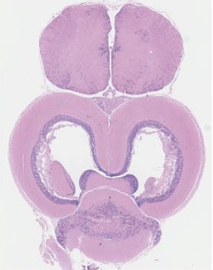
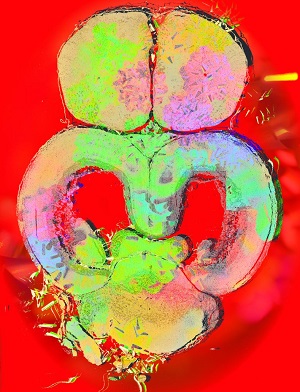
| Original Image | Artistic Reinterpretation The Mind that Knows Itself |
|
|---|---|---|
 |
 |
This image is a top view of an extremely thin slice of a fish brain. The colors are the result of staining; dense cell nuclei appear blue while proteins appear in shades of red. The frontal bulbs (located at the top of the image) form the telencephelon. This structure is most closely related to the amygdala in humans, and plays roles in memory, decision-making, and emotion. The larger, hollow middle lobes form the diencephelon, primarily responsible for vision. Finally, the central hind bulb is the cerebellum, which, as in humans, is responsible for coordinating movement
This particular image was taken from an experiment studying the best ways to preserve brain tissue for later study. H&E staining, like what was performed here, is the “gold standard” for tissue histology, useful for quickly visualizing physical features of the sample. Threespined stickleback - the fish species featured here - are studied in a number of research fields, including behavior and cognition, ecology, and evolution. Sectioning techniques, as used here, allow researchers to link behavioral genes to the locations in the brain where those genes are expressed. This is a key step toward unlocking the secrets of how genes and the brain control behavior.
Read local press coverage on the gallery event event here with my image featured.
See all the images on IGB’s facebook page.